nid: 59048
Additional formats:
None available
Description:
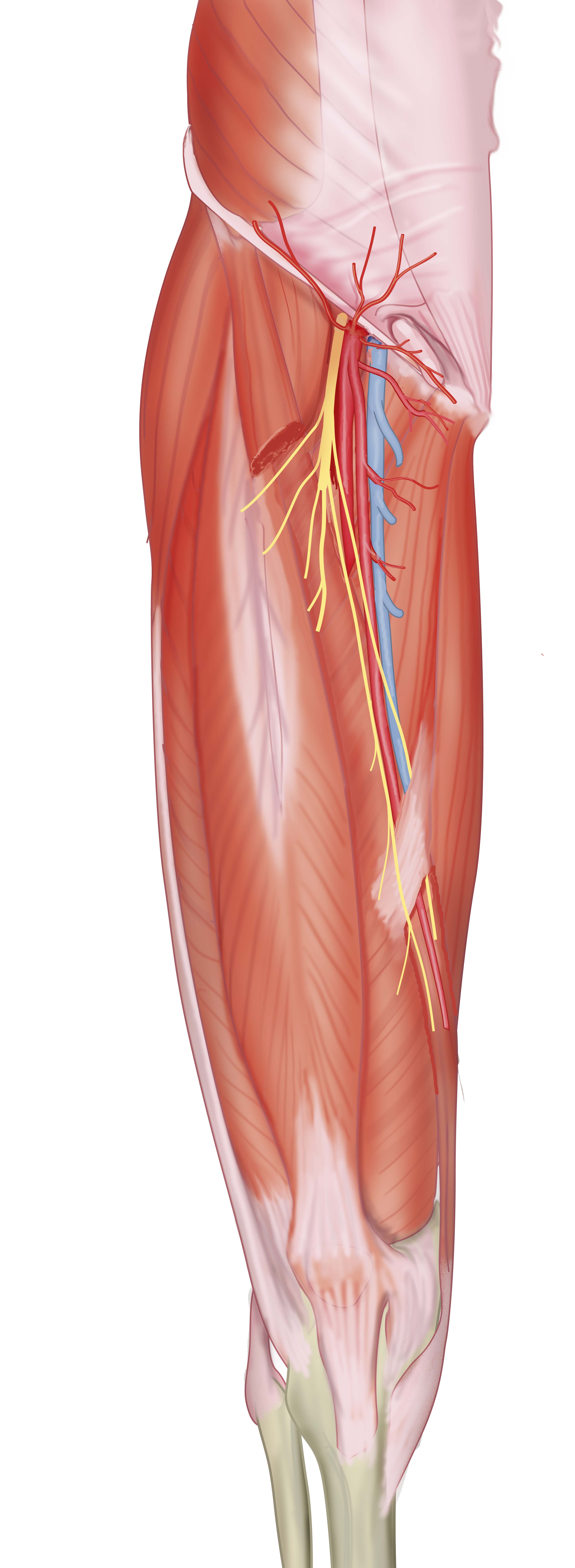
Anterior view of the thigh with femoral artery, vein and nerve. The sartorius muscle has been partially removed to show the anteromedial intermuscular septum. The origin of the deep femoral artery is also visible. No labels.
Illustration by Ron Slagter.
Illustration by Ron Slagter.
Anatomical structures in item:
Uploaded by: opgobee
Netherlands, Leiden – Leiden University Medical Center, Leiden University
Trunk of femoral artery
Arteria femoralis
Vena femoralis
Nervus femoralis
Femur
Arteria profunda femoris
Septum intermusculare vastoadductorium
Musculus quadratus femoris
Musculus rectus femoris
Musculus vastus lateralis
Musculus vastus medialis
Canalis adductorius
Hiatus adductorius
Musculus adductor longus
Musculus gracilis
Anulus inguinalis superficialis
Arteria circumflexa iliaca superficialis
Arteria epigastrica superficialis
Arteria pudenda externa superficialis
Arteria pudenda externa profunda
Ligamentum inguinale
Creator(s)/credit: Ron Slagter NZIMBI, medical illustrator, LUMC; Friso P. Jansen MD, anatomist, LUMC
Requirements for usage
You are free to use this item if you follow the requirements of the license:  View license
 View license
 If you use this item you should credit it as follows:
- For usage in print - copy and paste the line below:
- For digital usage (e.g. in PowerPoint, Impress, Word, Writer) - copy and paste the line below (optionally add the license icon):
"Slagter - Drawing Anterior thigh muscles with femoral artery, vein and nerve - no labels" at AnatomyTOOL.org by Ron Slagter, LUMC and Friso P. Jansen, LUMC, license: Creative Commons Attribution-NonCommercial-ShareAlike
"Slagter - Drawing Anterior thigh muscles with femoral artery, vein and nerve - no labels" by Ron Slagter, LUMC and Friso P. Jansen, LUMC, license: CC BY-NC-SA


Comments