
nid: 58741
Additional formats:
None available
Description:
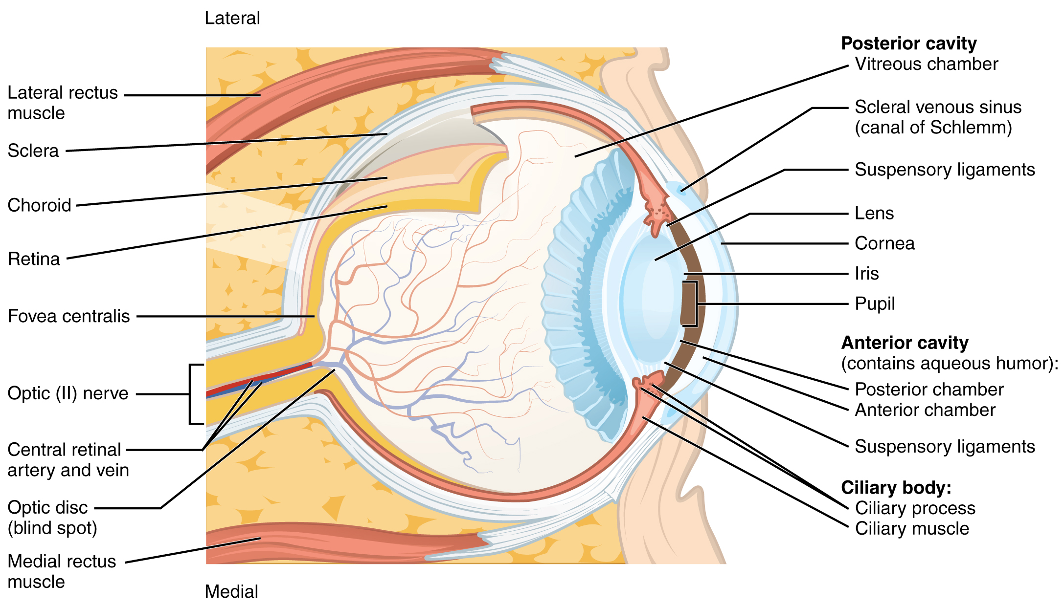
Structure of the Eye. The sphere of the eye can be divided into anterior and posterior chambers. The wall of the eye is composed of three layers: the fibrous tunic, vascular tunic, and neural tunic. Within the neural tunic is the retina, with three layers of cells and two synaptic layers in between. The center of the retina has a small indentation known as the fovea. English labels. From OpenStax book 'Anatomy and Physiology', fig. 14.15.
Anatomical structures in item:
Uploaded by: Jorn IJkhout
Netherlands, Leiden – Leiden University Medical Center, Leiden University
Oculus
Musculus rectus lateralis
Sclera
Choroidea
Retina
Fovea centralis
Nervus opticus
Arteria centralis retinae
Vena centralis retinae
Discus nervi optici
Musculus rectus medialis
Camera vitrea bulbi oculi
Sinus venosus sclerae
Zonula ciliaris
Lens
Cornea
Iris
Pupilla
Camera posterior bulbi oculi
Camera anterior bulbi oculi
Corpus ciliare
Processus ciliares
Musculus ciliaris
Creator(s)/credit: OpenStax
Requirements for usage
You are free to use this item if you follow the requirements of the license:  View license
View license If you use this item you should credit it as follows:
- For usage in print - copy and paste the line below:
- For digital usage (e.g. in PowerPoint, Impress, Word, Writer) - copy and paste the line below (optionally add the license icon):
"OpenStax AnatPhys fig.14.15 - Structure of the Eye - English labels" at AnatomyTOOL.org by OpenStax, license: Creative Commons Attribution. Source: book 'Anatomy and Physiology', https://openstax.org/details/books/anatomy-and-physiology.
"OpenStax AnatPhys fig.14.15 - Structure of the Eye - English labels" by OpenStax, license: CC BY. Source: book 'Anatomy and Physiology', https://openstax.org/details/books/anatomy-and-physiology.


Comments