
nid: 58680
Additional formats:
None available
Description:
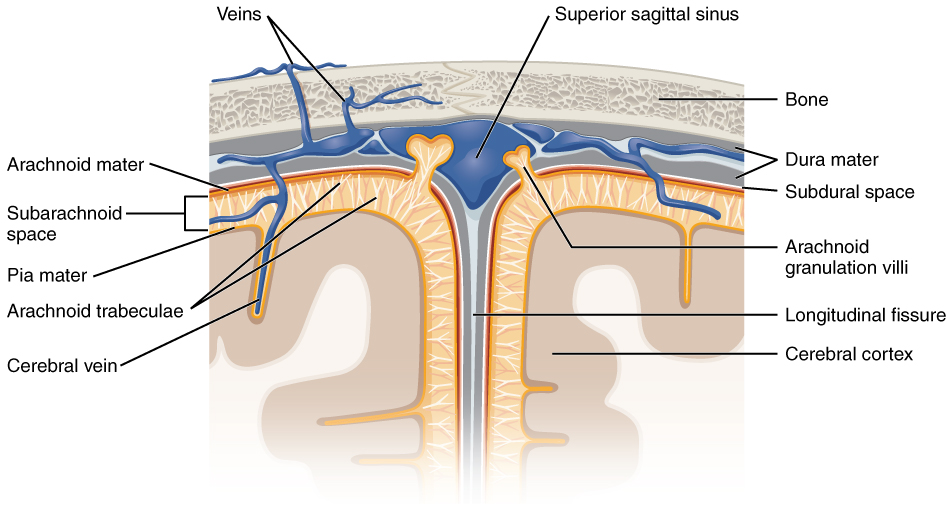
Meningeal Layers of Superior Sagittal Sinus. The layers of the meninges in the longitudinal fissure of the superior sagittal sinus are shown, with the dura mater adjacent to the inner surface of the cranium, the pia mater adjacent to the surface of the brain, and the arachnoid and subarachnoid space between them. An arachnoid villus is shown emerging into the dural sinus to allow CSF to filter back into the blood for drainage.
English labels. From OpenStax book 'Anatomy and Physiology', fig. 13.17.
English labels. From OpenStax book 'Anatomy and Physiology', fig. 13.17.
Anatomical structures in item:
Uploaded by: Jorn IJkhout
Netherlands, Leiden – Leiden University Medical Center, Leiden University
Encephalon
Arachnoidea mater
Pia mater
Trabeculae arachnoideae
Venae encephali
Sinus sagittalis superior
Dura mater
Spatium subdurale
Granulationes arachnoideae
Fissura longitudinalis cerebri
Pallium
Creator(s)/credit: OpenStax
Requirements for usage
You are free to use this item if you follow the requirements of the license:  View license
View license If you use this item you should credit it as follows:
- For usage in print - copy and paste the line below:
- For digital usage (e.g. in PowerPoint, Impress, Word, Writer) - copy and paste the line below (optionally add the license icon):
"OpenStax AnatPhys fig.13.17 - Meningeal Layers - English labels " at AnatomyTOOL.org by OpenStax, license: Creative Commons Attribution. Source: book 'Anatomy and Physiology', https://openstax.org/details/books/anatomy-and-physiology.
"OpenStax AnatPhys fig.13.17 - Meningeal Layers - English labels " by OpenStax, license: CC BY. Source: book 'Anatomy and Physiology', https://openstax.org/details/books/anatomy-and-physiology.


Comments