
nid: 58571
Additional formats:
None available
Description:
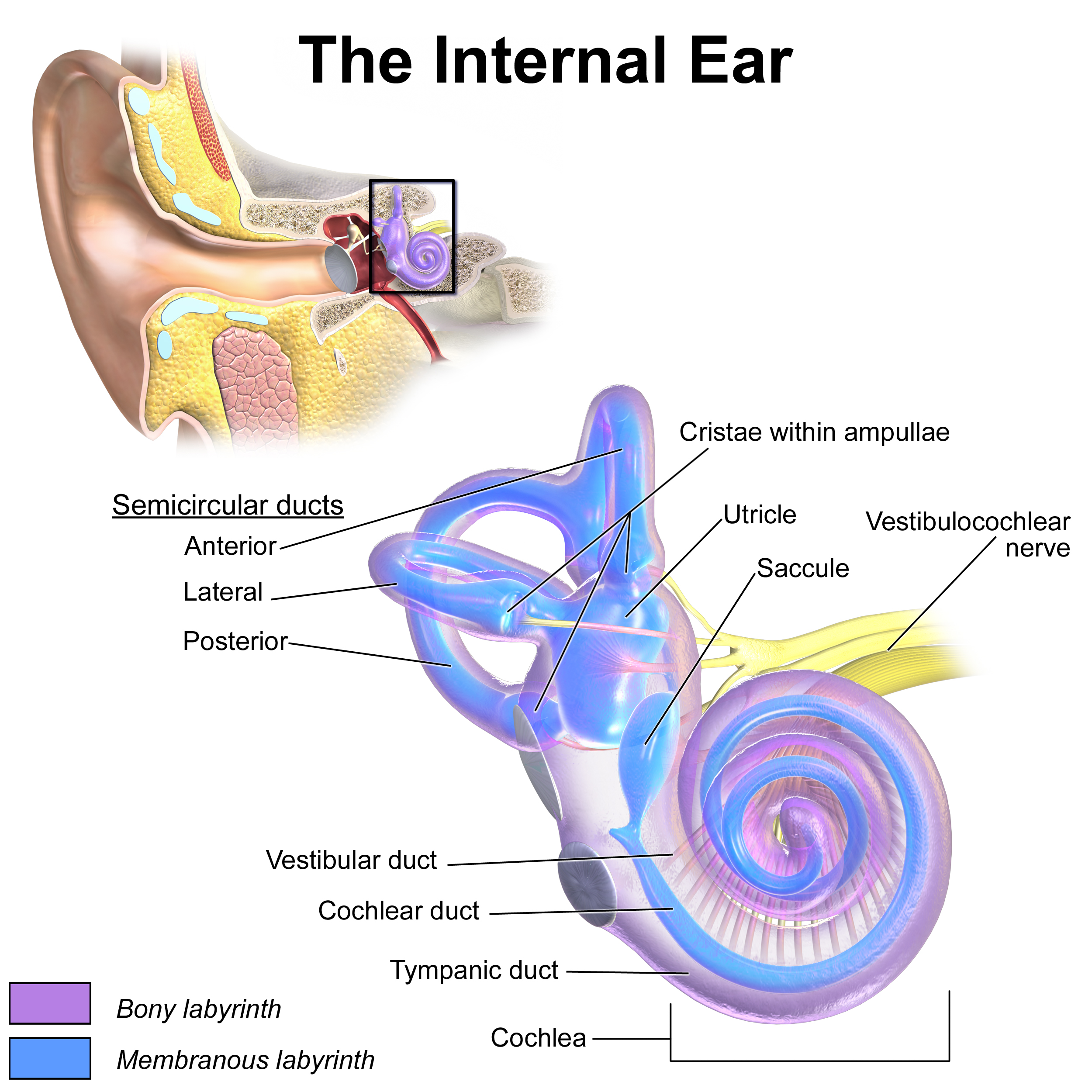
Anatomy of the internal ear. English labels.
By Blausen.com staff (2014). Retrieved from Medical gallery of Blausen Medical 2014., WikiJournal of Medicine 1 (2): 10. DOI:10.15347/wjm/2014.010. ISSN 2002-4436. Fig. 0329.
By Blausen.com staff (2014). Retrieved from Medical gallery of Blausen Medical 2014., WikiJournal of Medicine 1 (2): 10. DOI:10.15347/wjm/2014.010. ISSN 2002-4436. Fig. 0329.
Anatomical structures in item:
Uploaded by: Student10
Netherlands, Leiden – Leiden University Medical Center, Leiden University
Auris interna
Labyrinthus osseus
Labyrinthus membranaceus
Utriculus
Sacculus
Ductus semicirculares
Ductus semicircularis anterior
Cochlea
Ductus cochlearis
Ductus semicircularis posterior
Ductus semicircularis lateralis
Ampulla ossea lateralis
Ampulla ossea posterior
Ampulla ossea anterior
Nervus vestibularis
Nervus cochlearis
Scala tympani
Scala media
Scala vestibuli
Creator(s)/credit: Blausen.com staff (2014)
Requirements for usage
You are free to use this item if you follow the requirements of the license:  View license
View license If you use this item you should credit it as follows:
- For usage in print - copy and paste the line below:
- For digital usage (e.g. in PowerPoint, Impress, Word, Writer) - copy and paste the line below (optionally add the license icon):
"Blausen 0329 - Anatomy of the internal ear - English labels " at AnatomyTOOL.org by Blausen.com staff (2014), license: Creative Commons Attribution. Source: "Medical gallery of Blausen Medical 2014" https://en.wikiversity.org/wiki/WikiJournal_of_Medicine/Medical_gallery_of_Blausen_Medical_2014
"Blausen 0329 - Anatomy of the internal ear - English labels " by Blausen.com staff (2014), license: CC BY. Source: "Medical gallery of Blausen Medical 2014" https://en.wikiversity.org/wiki/WikiJournal_of_Medicine/Medical_gallery_of_Blausen_Medical_2014


Comments