
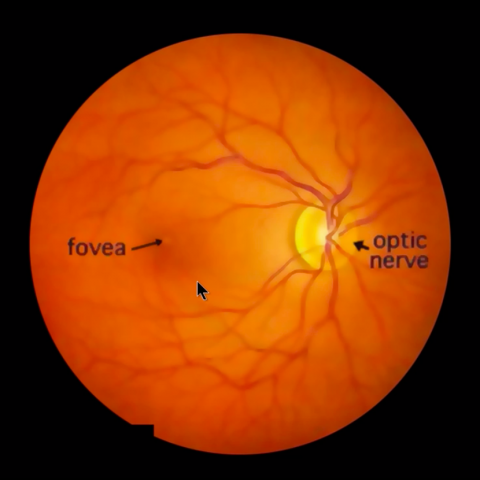
The Visual Pathway – Brain Dissections. The video briefly reviews the anatomy of the eye and the photic stimulation of the receptors is followed by a gross exploration of the visual pathway from the optic nerve, chiasm, and tract to the thalamus stressing how the left part of the visual world reaches the right hemisphere. Visual fields are related the retinotopic organization of the visual cortex. The eye as a window to the brain and its important vascular supply is also discussed. With English or Italian closed captions. Video by Suzanne S. Stensaas, PhD, Professor Emeritus, Department of Neurobiology and Anatomy, University of Utah School of Medicine. Video retrieved from https://neurologicexam.med.utah.edu/adult/html/brain-dissections.html#16
 View license
 If you use this item you should credit it as follows:
- For usage in print - copy and paste the line below:
- For digital usage (e.g. in PowerPoint, Impress, Word, Writer) - copy and paste the line below (optionally add the license icon):


Comments