
nid: 58441
Additional formats:
None available
Description:
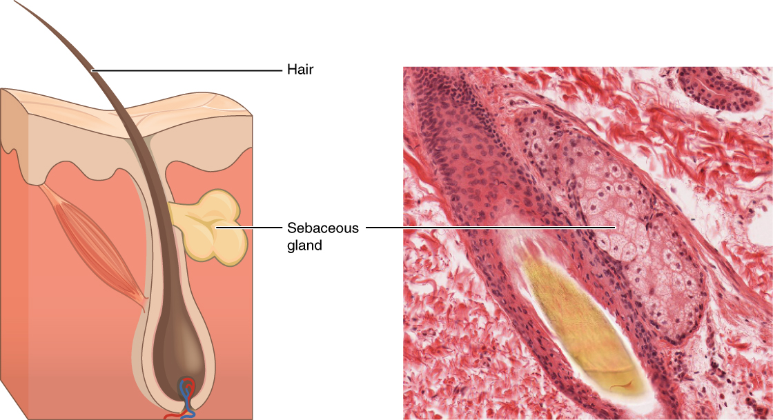
Sebaceous Glands. These glands secrete oils that lubricate and protect the skin. They are holocrine glands and they are destroyed after releasing their contents. New glandular cells form to replace the cells that are lost. LM × 400. (Micrograph provided by the Regents of University of Michigan Medical School © 2012). English labels. From OpenStax book 'Anatomy and Physiology', fig. 4.11.
Anatomical structures in item:
Uploaded by: Jorn IJkhout
Netherlands, Leiden – Leiden University Medical Center, Leiden University
Myocardium
Creator(s)/credit: OpenStax; Regents of U-M Medical School, UMich MedSchool
Requirements for usage
You are free to use this item if you follow the requirements of the license:  View license
View license If you use this item you should credit it as follows:
- For usage in print - copy and paste the line below:
- For digital usage (e.g. in PowerPoint, Impress, Word, Writer) - copy and paste the line below (optionally add the license icon):
"OpenStax AnatPhys fig.4.11 - Sebaceous Glands - English labels" at AnatomyTOOL.org by OpenStax and Regents of U-M Medical School, UMich MedSchool, license: Creative Commons Attribution. Source: book 'Anatomy and Physiology', https://openstax.org/details/books/anatomy-and-physiology.
"OpenStax AnatPhys fig.4.11 - Sebaceous Glands - English labels" by OpenStax and Regents of U-M Medical School, UMich MedSchool, license: CC BY. Source: book 'Anatomy and Physiology', https://openstax.org/details/books/anatomy-and-physiology.


Comments