
nid: 58421
Additional formats:
None available
Description:
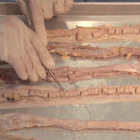
The Spinal Cord & Monosynaptic Reflex – The video demonstrates the spinal cord's relationship to the foramina, discs and spinal nerves. The dura, ganglia, and rootlets are shown as well as the gray and white matter in gross sections at different levels in a model and gross sections. Also shown and described is the anatomy of a monosynaptic reflex and the concept of a dermatome. With English or Italian closed captions. Video by Suzanne S. Stensaas, PhD, Professor Emeritus, Department of Neurobiology and Anatomy, University of Utah School of Medicine. Video retrieved from: https://neurologicexam.med.utah.edu/adult/html/brain-dissections.html#06
Anatomical structures in item:
Uploaded by: M_Orsatti
Netherlands, Leiden – Leiden University Medical Center, Leiden University
Vertebra
Medulla spinalis
Columna vertebralis
Cauda equina
Pars duralis fili terminalis
Os sacrum [vertebrae sacrales I - V]
Discus intervertebralis
Canalis centralis medullae spinalis
Ganglion vertebrale
Dorsal root
Radix anterior (Nervus spinalis)
Radix anterior (Nervus spinalis)
Radix posterior (Nervus spinalis)
Arteria basilaris
Arteria vertebralis
Creator(s)/credit: Professor Emeritus Suzanne S. Stensaas PhD
Requirements for usage
You are free to use this item if you follow the requirements of the license:  View license
View license If you use this item you should credit it as follows:
- For usage in print - copy and paste the line below:
- For digital usage (e.g. in PowerPoint, Impress, Word, Writer) - copy and paste the line below (optionally add the license icon):
"Utah NALab 06 - Brain Dissection Video: The Spinal Cord & Monosynaptic Reflex. Closed Captions in English and Italian " at AnatomyTOOL.org by Suzanne S. Stensaas, license: Creative Commons Attribution-NonCommercial-ShareAlike


Comments