
nid: 58343
Additional formats:
None available
Description:
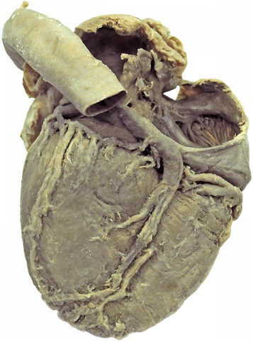
Posterior view of the heart. In this figure, an posterior view of the heart is visible with opened atria, so the anatomy of the ventral wall can be seen. With the buttons on the right you can light up different anatomic structures.
Anatomical structures in item:
Uploaded by: rva
Netherlands, Leiden – Leiden University Medical Center, Leiden University
Ramus circumflexus (Arteria coronaria sinistra)
Aorta
Sinus coronarius
Arteria coronaria dextra
Atrium sinistrum
Atrium dextrum
Vena cardiaca media
Ramus posterior ventriculi sinistri arteria coronariae sinistrae
Ventriculus dexter
Arteria coronaria dextra
Creator(s)/credit: Dr Claudia Krebs, UBC; Monika Fejtek, UBC; Alexa Mordhorst, UBC
Requirements for usage
You are free to use this item if you follow the requirements of the license:  View license
View license If you use this item you should credit it as follows:
- For usage in print - copy and paste the line below:
- For digital usage (e.g. in PowerPoint, Impress, Word, Writer) - copy and paste the line below (optionally add the license icon):
"U.Br.Columbia ClinAnat - Interactive Page posterior view of a plastinated heart" at AnatomyTOOL.org by Claudia Krebs, UBC, Monika Fejtek, UBC and Alexa Mordhorst, UBC, license: Creative Commons Attribution-NonCommercial-ShareAlike
"U.Br.Columbia ClinAnat - Interactive Page posterior view of a plastinated heart" by Claudia Krebs, UBC, Monika Fejtek, UBC and Alexa Mordhorst, UBC, license: CC BY-NC-SA


Comments