
nid: 57867
Additional formats:
None available
Description:
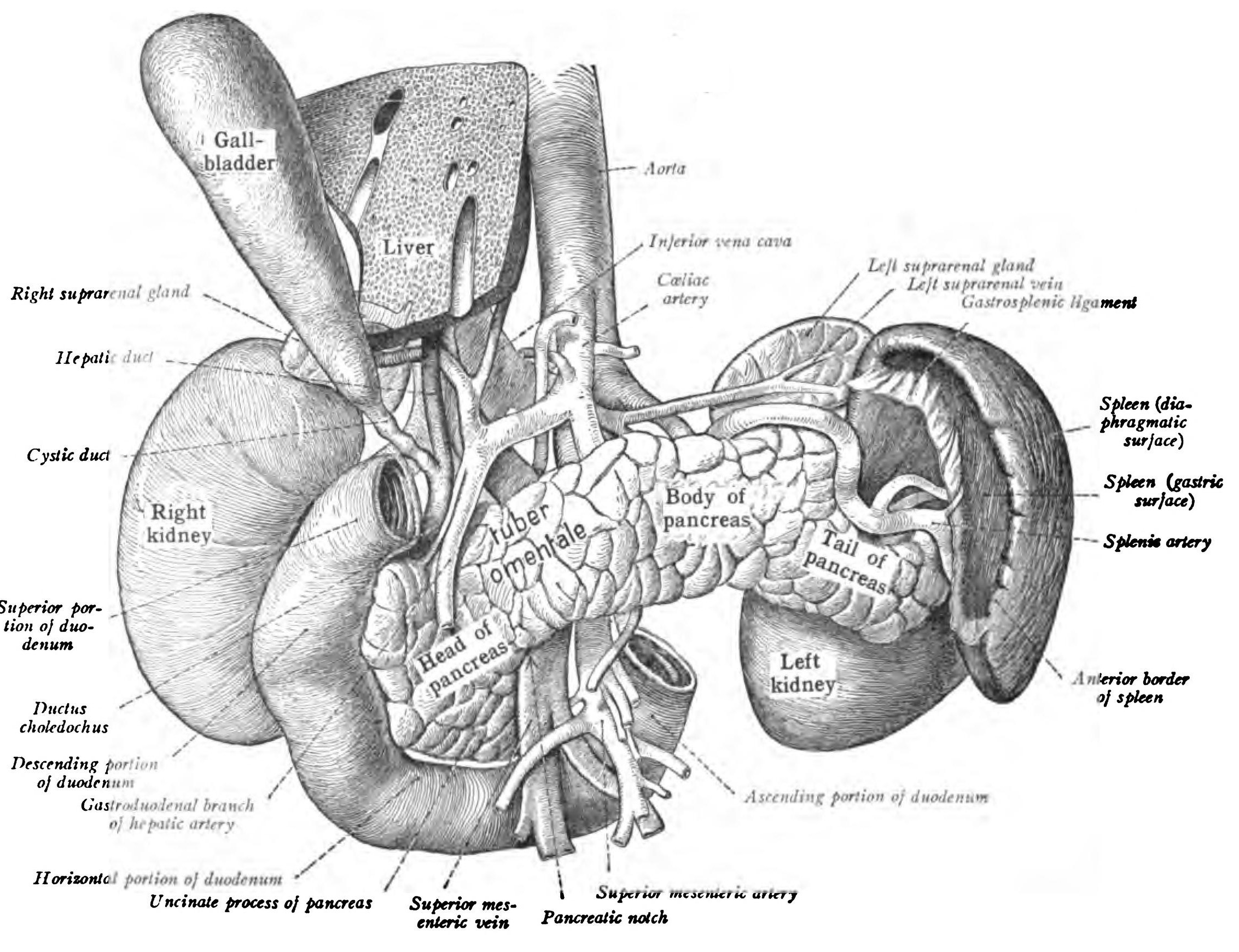
Duodenum, pancreas, spleen, kidney, suprarenal glands, gall bladder, aorta and vena cava: organs shown in their relative positions. English labels.
From 'Atlas and Textbook of Human Anatomy', 1906, Vol. 2, fig.394, by Johannes Sobotta and J. Playfair McMurrich. Artist: K. Hajek. Retrieved from Sobotta's Anatomy plates at Wikimedia. Possible original source: Sobotta's atlas at archive.org.
From 'Atlas and Textbook of Human Anatomy', 1906, Vol. 2, fig.394, by Johannes Sobotta and J. Playfair McMurrich. Artist: K. Hajek. Retrieved from Sobotta's Anatomy plates at Wikimedia. Possible original source: Sobotta's atlas at archive.org.
Anatomical structures in item:
Uploaded by: Student128
Netherlands, Leiden – Leiden University Medical Center, Leiden University
Pancreas
Duodenum
Ren (Nephros)
Glandula suprarenalis
Aorta
Vena cava inferior
Vesica biliaris (Fellea)
Hepar
Splen
Arteria lienalis
Vena suprarenalis sinistra
Truncus coeliacus
Ductus cysticus
Ductus hepaticus communis
Ductus biliaris
Creator(s)/credit: Prof.dr. Johannes Sobotta, anatomist
Requirements for usage
You are free to use this item.  Read more
Read more This item is in the Public Domain because its copyright has expired. You are not required to credit its creators when you use it. Nevertheless, it is adviced to do so. First, it is academically correct to pay tribute to the creators. Second, items of unknown origin might be classified as 'copyright infringement' by copyright controlling bodies, with possible resulting bills. Stating the item's source will prevent this. You can use the following text:
- For usage in print - copy and paste the line below:
- For digital usage (e.g. in PowerPoint, Impress, Word, Writer) - copy and paste the line below (optionally add the icon):
"Sobotta 1906 fig.394 - Duodenum, pancreas, spleen, kidney, suprarenal glands, gall bladder, aorta and vena cava - English labels" at AnatomyTOOL.org by Johannes Sobotta is in the Public Domain.


Comments