Project Open Anatomy Learning Materials (TOOL2)
In this project (September 2018- Sept 2020 [extended until Mid 2021]) a substantial scaling up in content will be achieved by a newly received grant for the development and disclosure of anatomical drawings, 3D models, embryology and clinical anatomical learning materials (link: in Dutch). Various Dutch (AUMC, LUMC, UM, UMCG) and Flemish universities (Ghent, Hasselt, Leuven) collaborate in this project. Furthermore, this project is supported by the NAV (Dutch Association of Anatomists). The grant was received from the Dutch Ministry of Education, Culture and Science, in its Open Online Education Incentive scheme.
For information about the original TOOL project please see here.
The project's three main goals are:
- Developing and/or opening up a significant amount of open anatomical learning materials and offering these on AnatomyTOOL for free use for anyone.
- Implementing these learning materials in regular education of the anatomical departments of the Universities (of Applied Sciences) in the Netherlands and Flanders.
- Growing a community of anatomists (co)creating, exchanging, using, sharing, reviewing and maintaining these open learning materials.
The project consists of 7 work packages, with as main content:
| 1. Organisation | |
|
|
2. General anatomy learning materials
Existing online open anatomical learning material will be inventoried, reviewed and offered on AnatomyTOOL. The aim is to come to direct links to quality open online images for a large part of the anatomical structures in education. Medical illustrator Ron Slagter, illustrator of the Nederlands Tijdschrift voor Geneeskunde (Netherlands Journal of Medicine) for many years, and illustrator for various medical projects, made, supervised by medical professionals and/or anatomists, hundreds of high quality illustrations, covering a large part of the anatomy. He is willing to release the illustrations under Creative Commons license on AnatomyTOOL. In this project they will be selected, prepared and published for this purpose. |
 |
3. 3D adult anatomy learning materials
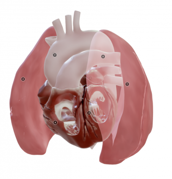
In this package, 3D models and/or derived visualisations such as 2D shots, and (stereoscopic) rotatable 3D videos, will be developed. The materials will be viewable in a regular web browser. The topics will probably be: the heart, pelvis and muscles. |
|
|
4. Embryology learning materials
3D embryo models and short educational clips based on these will be developed. These educational clips will show, per organ or anatomical structure, how the organ looks in a 3D model, and discuss how embryological development leads to the adult configuration, as well as how it may possibly result in congenital abnormalities and variations. |
 |
5. Anatomy in the clinic - learning materials
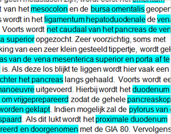
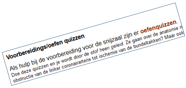
Readers will be made about anatomy during operations, on the basis of surgical protocols, with associated questions. Medical interns will be able to use these to prepare for operations they attend, to quickly and specifically retrieve the relevant anatomy. Clinical anatomical imaging materials will also be made available, e.g. 3D videos based on clinical MRIs / CTs, as well as surgical images. |
 |
6. Application in education and evaluation
The developed materials will be used in education. In this work package, the experiences of teachers and students with this usage will be evaluated. |
 |
7 Libraries and open learning platforms
This work package will investigate, together with the scientific libraries (LUMC, UM, AUMC, UMCG) to what extent libraries can play a structural role in an open learning platform. They will support in the inventory of sources and in researching the best ways of (automated) metadation and of reviewing. Training will also be developed for teachers about Creative Commons licenses and about searching for and using open material. |
More information about the TOOL project: contact Paul Gobée (o.p.gobee@lumc.nl)
- 52325 reads
