Region
System
Collections
Embryology
Microscopy
Dissections
Radiology
Clinical Anatomy
Documents
Images
Videos
3D models, games
Lessons. quizzes
Viewers
Create & edit content
Take quiz
Create account
/
Forgot password
About
Project Open Anatomy Learning Materials (TOOL2)
Manuals
Anatomy Learning Resources
Credits
Medical disclaimer
Legal information
Donate
Sponsor
Contact
Open 3D Model
Archive
Reviewers - areas of interest anatomists
Information
Learn
Search:
Subject
Technique
View
- Any -
Patient seen from the front
Patient seen from the front left
Patient seen from the left
Patient seen from the back left
Patient seen from the back
Patient seen from the back right
Patient seen from the right
Patient seen from the front right
Patient supine, viewed from the feet
Patient supine, viewed from the feet / left
Patient supine, viewed from the left
Patient supine, viewed from the head / left
Patient supine, viewed from the head
Patient supine, viewed from the head / right
Patient supine, viewed from the right
Patient supine, viewed from the feet / right
Patient prone (lying face down or on her side), viewed from the head, the side, or the feet
Other view direction or unknown
System
Body region
Sobotta 1906 fig.401 - Third stage of development of the intestinal canal and peritoneum - English labels
Image:
Sobotta 1906 fig.402 - Formation of the greater omentum - English labels
Image:
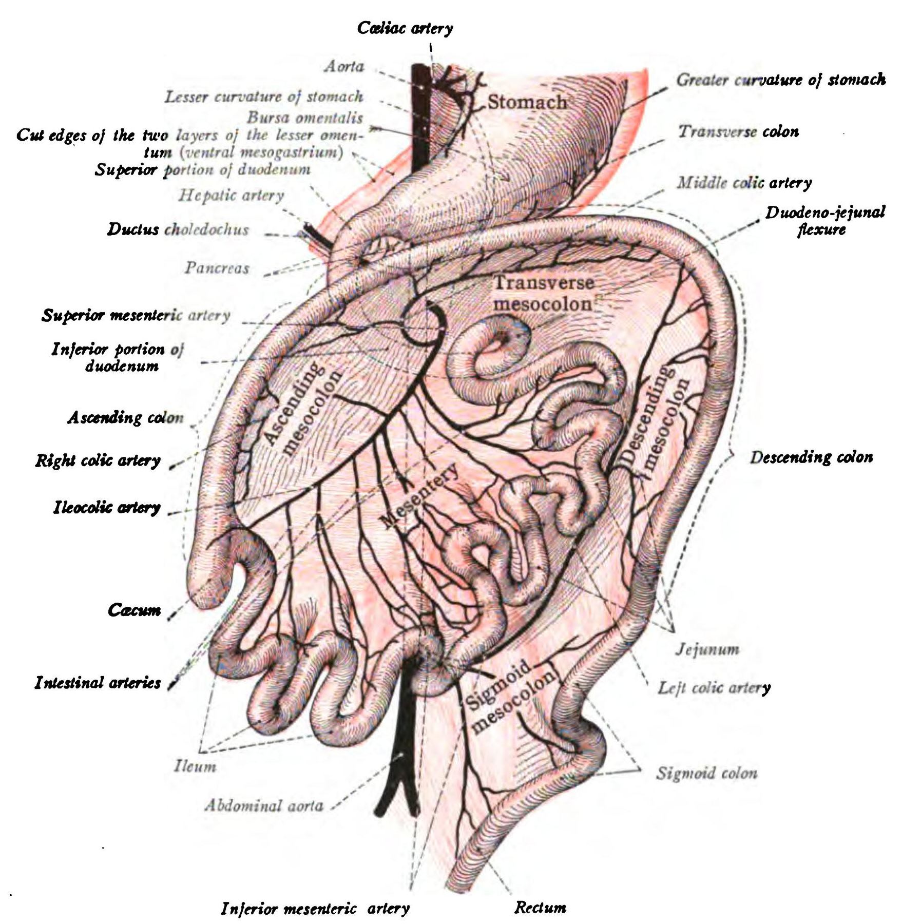
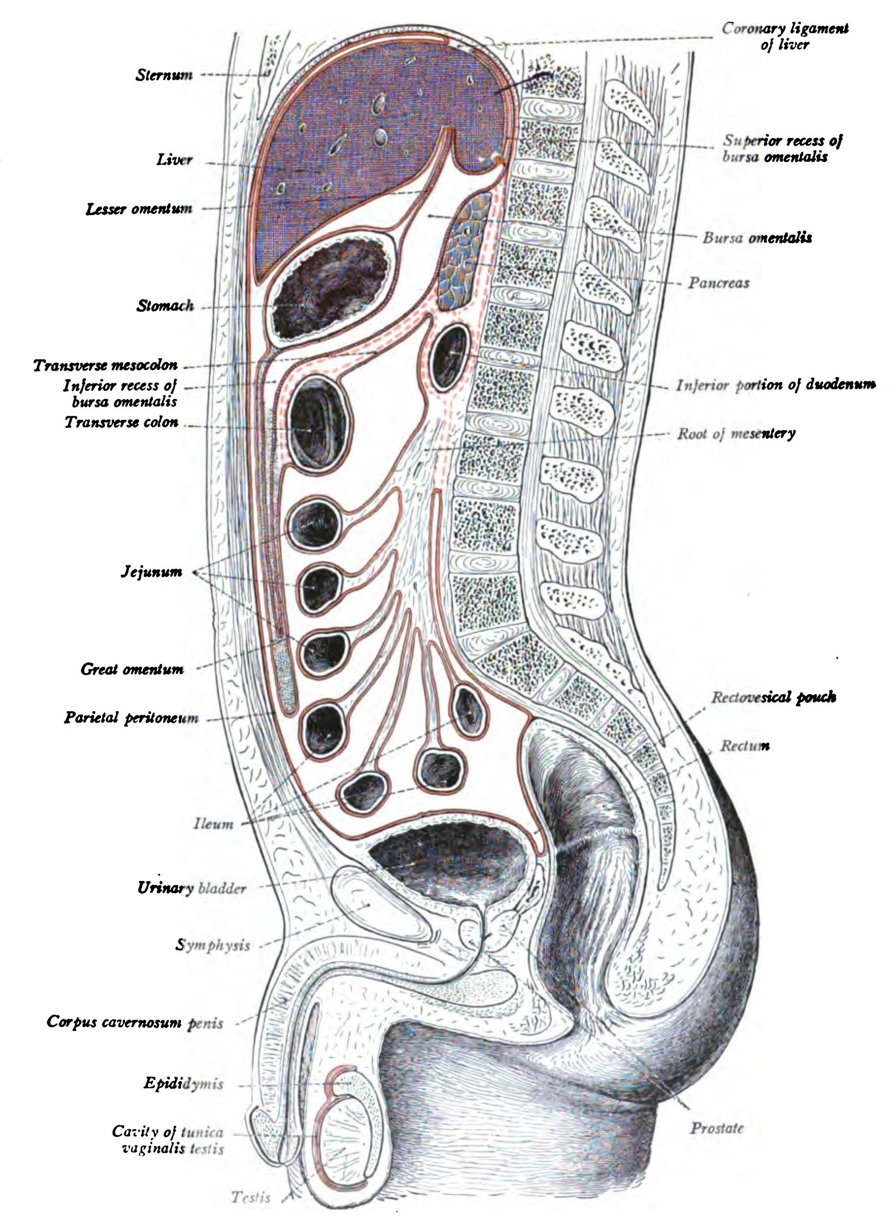
Sobotta 1906 fig.403 - Peritoneal arrangement - English labels
Image:
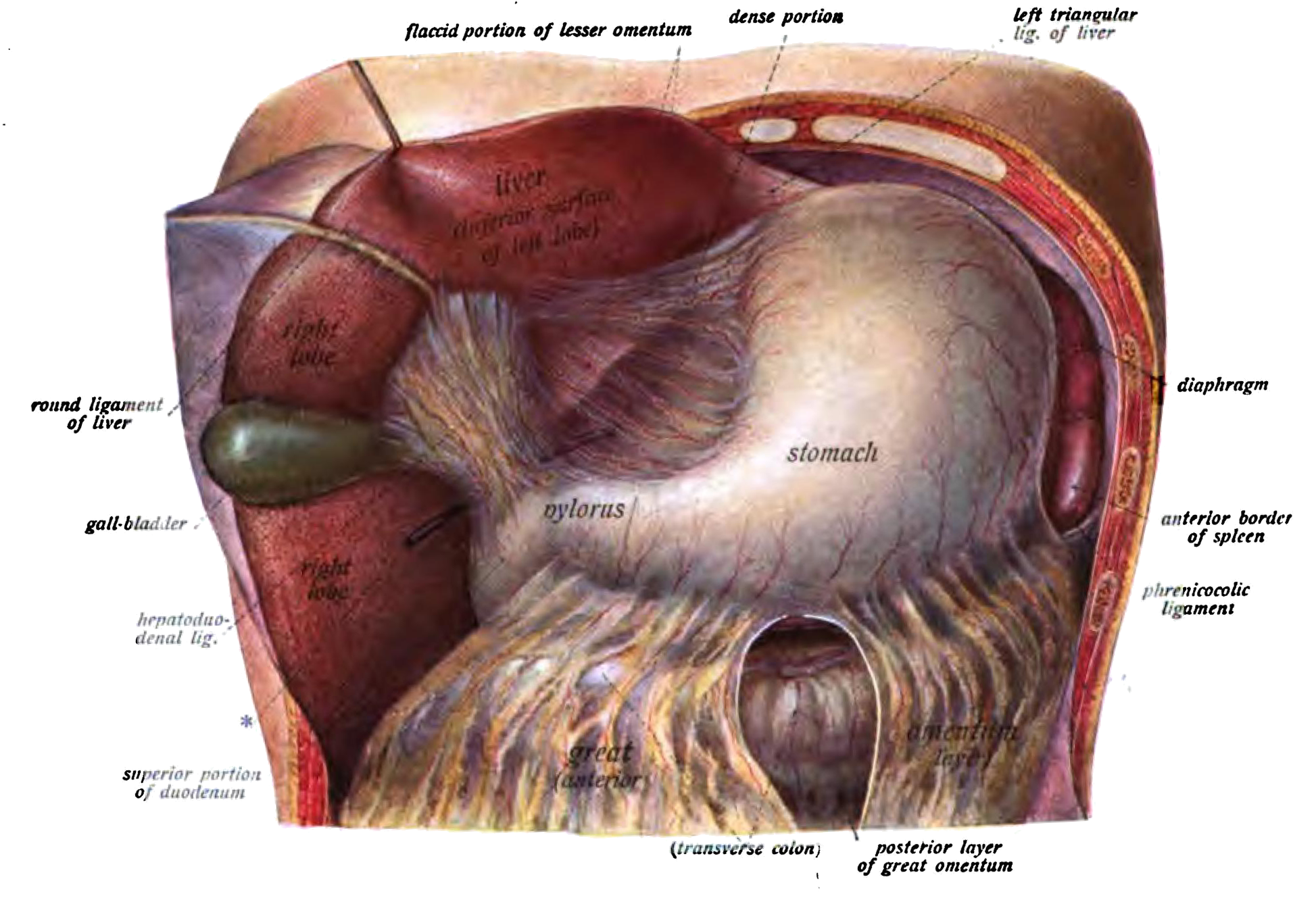
Sobotta 1906 fig.405 - Upper portion of the abdominal cavity - English labels
Image:
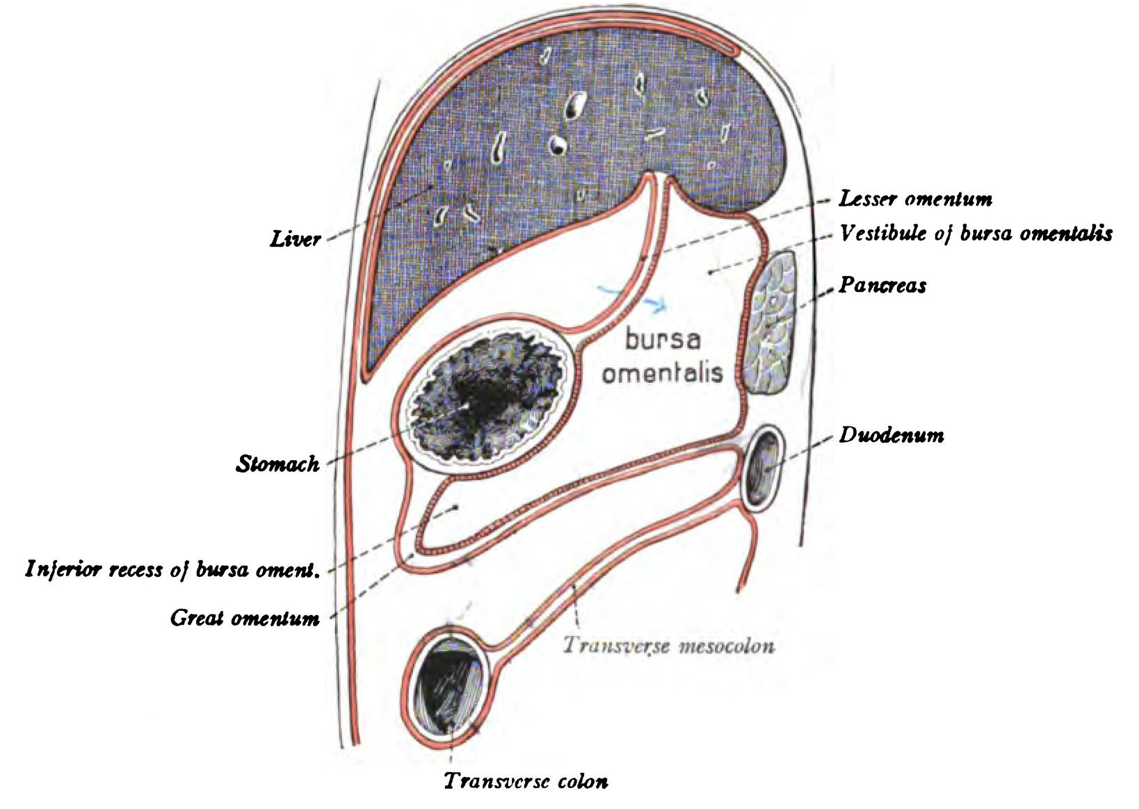
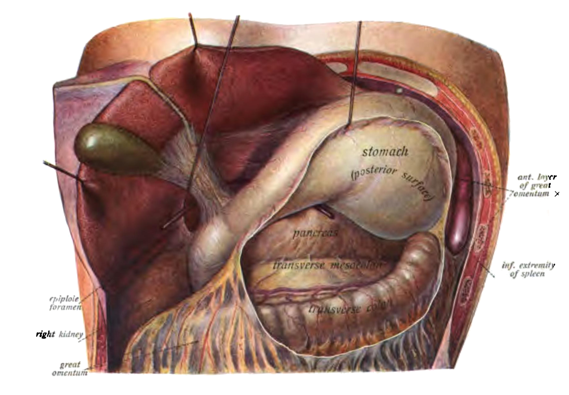
Sobotta 1906 fig.406 - Epiploic foramen and bursa omentalis - English labels
Image:
Sobotta 1906 fig.407 - Omentum majus - English labels
Image:
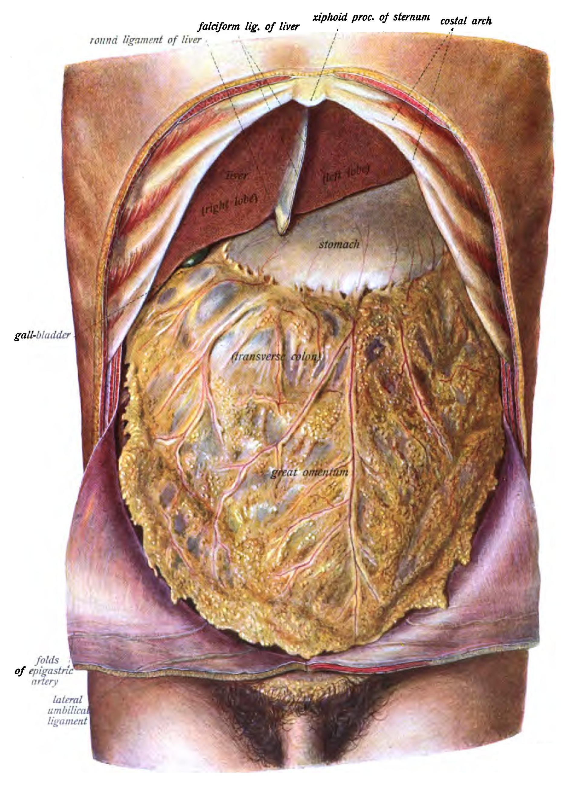
Sobotta 1906 fig.408 - The abdominal viscera - English labels
Image:
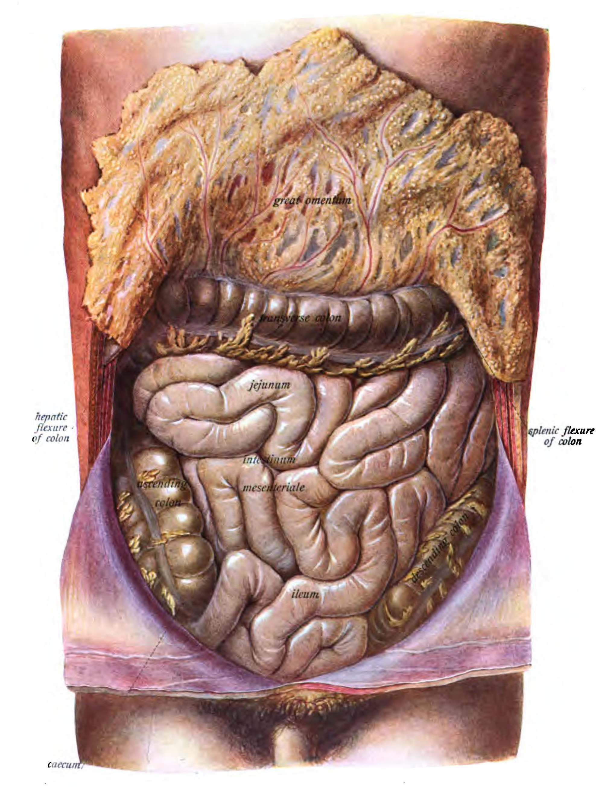
Sobotta 1906 fig.409 - The abdominal viscera, small intestine removed - English labels
Image:
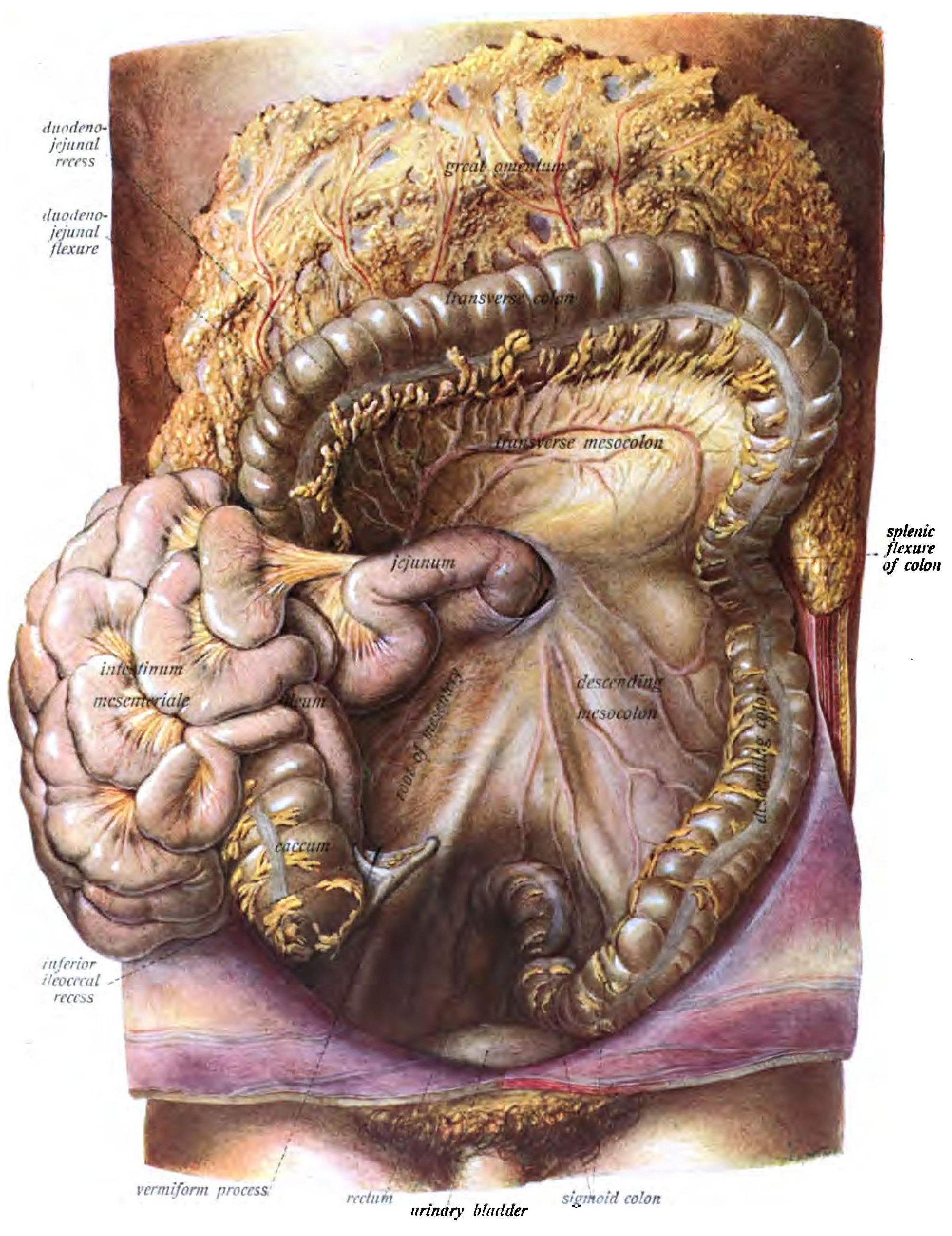
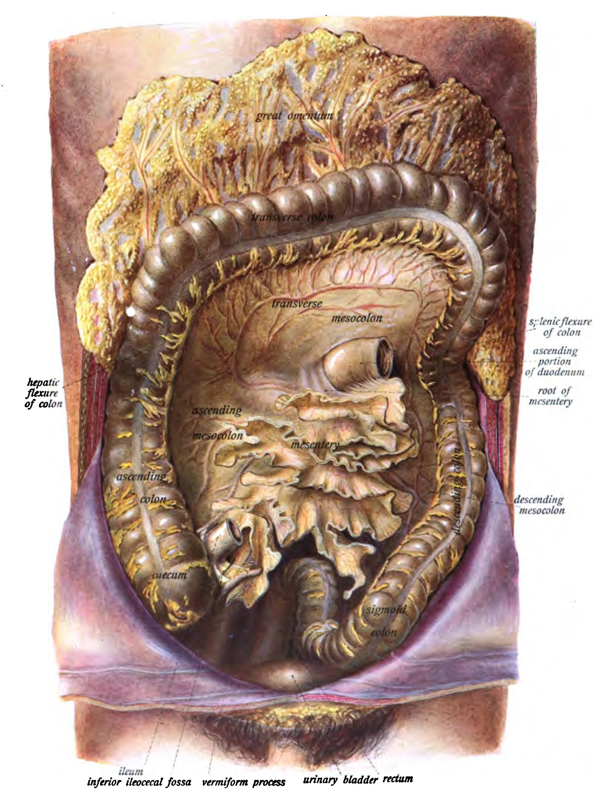
Sobotta 1906 fig.410 - Colon in situ - English labels
Image:
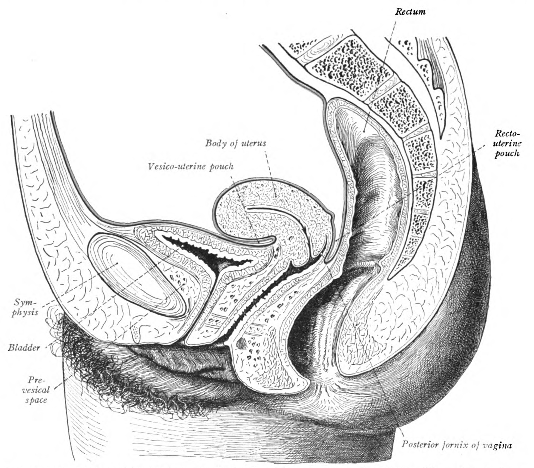
Sobotta 1906 fig.411 - Peritoneal arrangement in the female pelvis - English labels
Image:
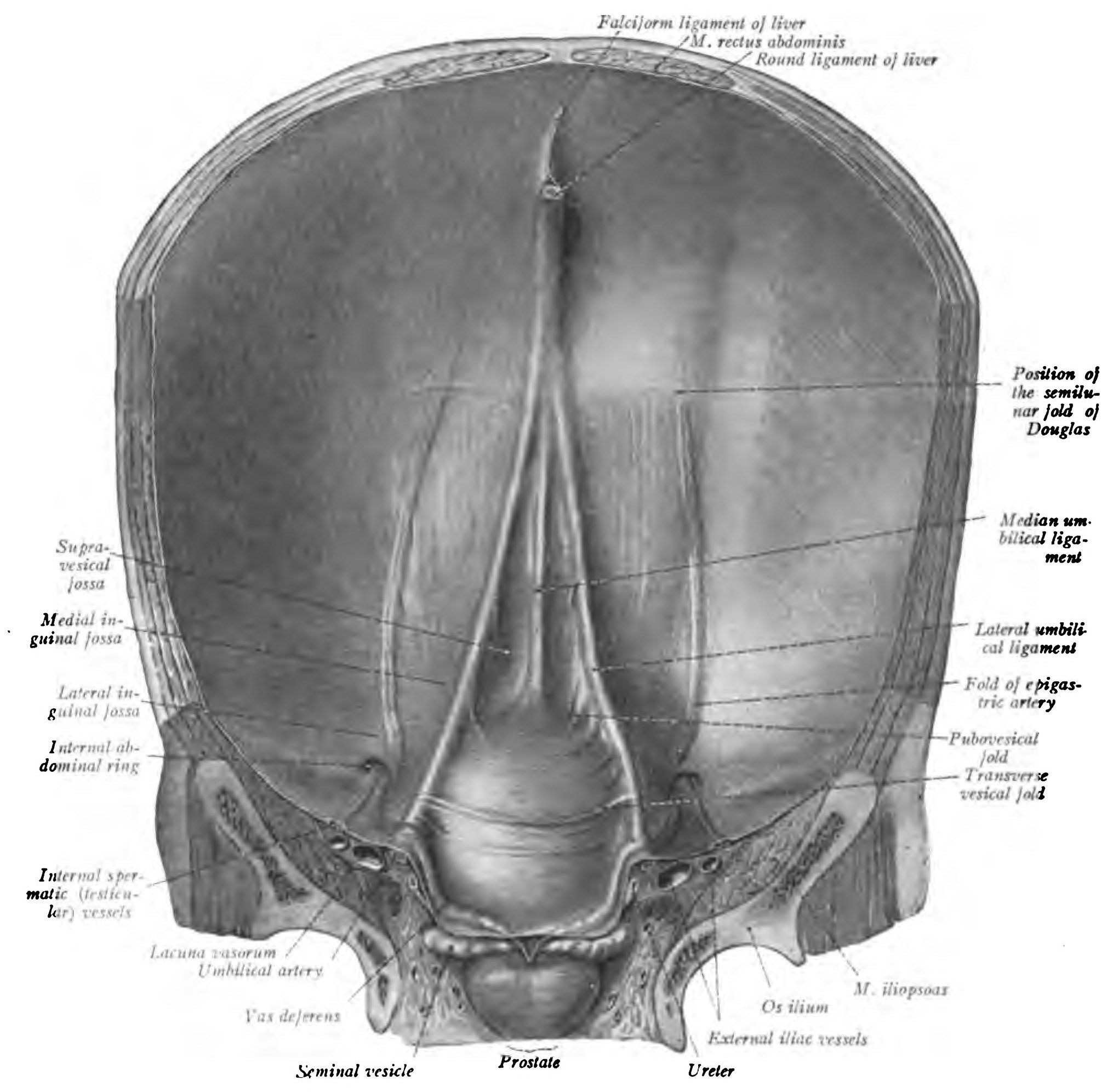
Sobotta 1906 fig.412 - Anterior abdominal wall of a newborn - English labels
Image:
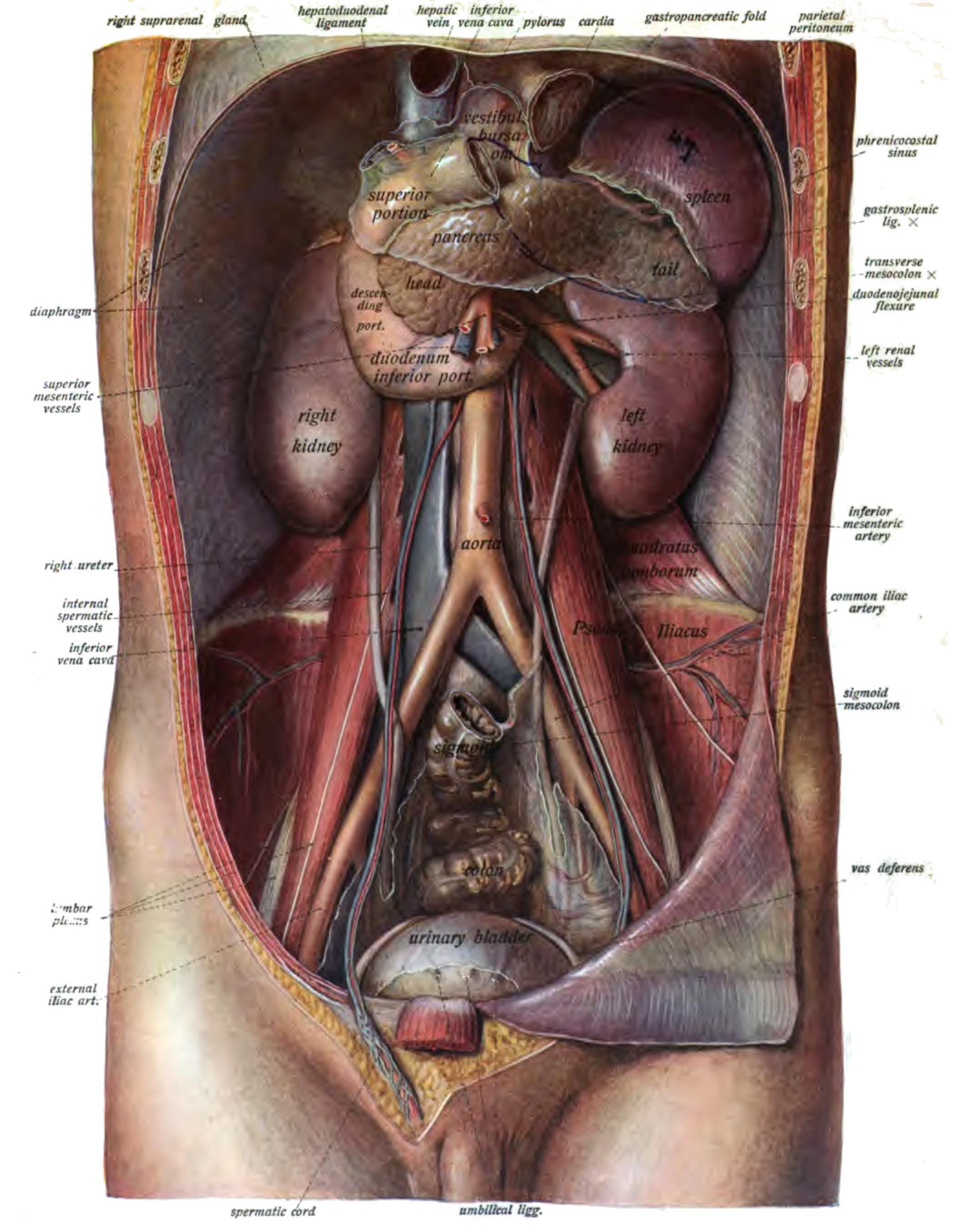
Sobotta 1906 fig.413 - Viscera on the posterior abdominal wall - English labels
Image:
Pages
« first
‹ previous
…
43
44
45
46
47
48
49
50
51
…
next ›
last »